
- Svi članci
- Dijagnostika
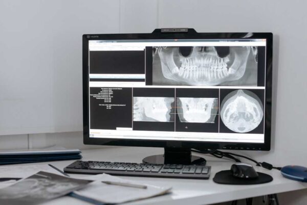
Jedna od najpopularnijih usluga u Medikadentu je naša linija CBCT snimanja, što je kratica za kompjutoriziranu tomografiju s konusnim snopom....
Jedna od najpopularnijih usluga u Medikadentu je naša linija CBCT snimanja, što je kratica za kompjutoriziranu tomografiju s konusnim snopom....